

MRI and CT scans are essential tools in detecting and diagnosing sarcoma. MRI provides detailed soft tissue imaging, helping doctors assess the tumor’s size, location, and extent. It uses strong magnetic fields and radio waves to produce high-resolution images.
CT scans offer cross-sectional views, revealing the tumor’s size, shape, and relationship to surrounding structures. They use X-ray technology combined with computer processing to provide detailed information.
MRI and CT scans enable accurate sarcoma diagnosis, guiding treatment decisions and improving patient outcomes.
Understanding MRI (Magnetic Resonance Imaging) Scans For Sarcoma Diagnosis
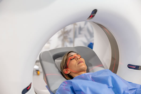
Magnetic Resonance Imaging (MRI) is a noninvasive technique that creates detailed images using magnetic fields and radio waves. It is especially valuable for diagnosing sarcoma, a cancer of connective tissues. Patients lie on a table during an MRI, and the machine generates images using magnetic fields and radio waves. Unlike other imaging methods, MRI doesn’t use ionizing radiation, making it safer for repeated use in monitoring sarcoma progression.
MRI is particularly effective in differentiating sarcoma from other soft tissue masses. Its high contrast resolution helps oncologists assess the tumor’s nature and plan treatments.
The Role Of CT (Computed Tomography) Scans In Detecting Sarcoma
CT scans are crucial in diagnosing sarcoma, providing cross-sectional images through X-ray technology and computer processing. While MRI is superior for soft tissue imaging, CT scans, according to Tellica Imaging, excel at visualizing bony structures and internal organs, offering a comprehensive view of the tumor’s relationship with surrounding anatomy. During the scan, patients move through a scanner while X-ray images are taken from different angles, enabling the computer to create detailed views of the tumor’s size, shape, and density. Often used alongside MRI, CT scans help assess metastasis stage sarcoma and guide treatment decisions.
Advantages And Limitations Of MRI Scans For Sarcoma Imaging
MRI scans offer significant benefits in sarcoma imaging, particularly with exceptional soft tissue contrast that helps visualize tumors, their margins, and their relationships with surrounding tissues. This aids in surgical planning and understanding tumor extent. MRI is also radiation-free, making it safer for patients needing multiple scans.
However, MRI has limitations, including higher cost, longer scan duration, and limited accessibility compared to CT. Certain implants or devices may also prevent patients from undergoing an MRI.
Advantages And Limitations Of CT Scans For Sarcoma Imaging
CT scans offer several benefits for imaging sarcoma. They are quick and convenient, particularly for patients with difficulty staying still for extended periods. CT scans are also valuable for evaluating bony structures and detecting metastasis, making them crucial in treatment planning and prognosis.
However, CT scans involve ionizing radiation, which can pose risks with repeated use, especially in younger patients or those requiring long-term monitoring. While they provide excellent spatial resolution, they may not differentiate soft tissue types as effectively as MRI, occasionally complicating diagnosis.
Key Differences Between MRI and CT Scans In Sarcoma Detection
Understanding the differences between MRI and CT scans is crucial for healthcare providers and patients. MRI uses magnetic fields and radio waves, excelling in soft tissue contrast. This makes it ideal for visualizing sarcomas and aiding surgical planning. In contrast, CT scans use X-rays and are better for assessing bony structures and detecting metastasis, which is vital for cancer staging.
MRI typically requires longer patient immobilization, while CT scans are quicker and less stressful. Each method has specific contraindications, so healthcare providers must choose the most suitable imaging technique based on the patient’s needs.
How MRI And CT Scans Complement Each Other In Sarcoma Diagnosis
MRI and CT scans complement each other in diagnosing sarcoma, providing a more complete understanding of the tumor. A CT scan assesses overall anatomy and identifies metastases, while an MRI offers detailed images of the tumor’s relationship with surrounding tissues, refining the diagnosis and guiding treatment.
Using both techniques also improves the monitoring of treatment responses and recurrence detection. MRI shows soft tissue changes, while CT scans assess anatomical shifts, ensuring timely identification of changes in tumor status. This combined approach enhances modern sarcoma care and improves patient outcomes.
Preparing For An MRI or CT scan For Sarcoma Detection
MRI and CT scan preparation can vary by procedure and facility. Patients should wear metal-free clothing for an MRI, and sometimes, a contrast agent is used, requiring specific food and drink instructions. Patients may need to fast for CT scans if contrast material is used, and allergies to iodine-based agents must be disclosed. Sharing medical history, including surgeries or implants, is essential for selecting the appropriate imaging method. The Sarcoma Oncology Center emphasizes proper imaging preparation for accurate sarcoma cancer diagnosis and treatment planning.
Interpreting MRI and CT Scan Results For Sarcoma Diagnosis
Interpreting MRI and CT scans involves assessing the tumor’s size, location, and characteristics to help diagnose sarcoma. In MRI, radiologists focus on the tumor’s shape, margins, and signal intensity to differentiate between benign and malignant masses. CT scans evaluate the tumor’s density and morphology to detect malignancy and metastasis.
These imaging results are combined with clinical and lab findings to guide treatment decisions, improving patient outcomes.
Conclusion
MRI and CT scans are essential for accurately detecting sarcoma. Each modality provides unique insights into the tumor’s characteristics, extent, and relationship with surrounding structures. This combined approach is crucial for developing effective treatment strategies and improving patient care.
By utilizing both imaging techniques, healthcare providers can ensure timely and accurate diagnosis, enhancing sarcoma management and improving patient outcomes and quality of life.
With ongoing advancements in imaging technology, the future of sarcoma detection and treatment holds great promise, offering more targeted and effective therapies for patients.


Comments